Introduction: What people really want from plant disease identification
When someone searches for plant disease identification they want three things: to identify the problem quickly, to know if it’s contagious or dangerous, and to get clear next steps to treat or prevent it. Accurate identification reduces unnecessary sprays, saves plants, and prevents spread. This guide translates field mycology, plant pathology, and practical horticulture into a systematic approach you can use for houseplants, vegetable gardens, ornamentals and trees.
- Quick visual cues for common pathogens
- Step-by-step diagnostic workflow you can follow with your phone
- Practical treatment, prevention, and safety guidance
Why accurate plant disease identification matters
Misdiagnosis can make problems worse. For example, applying a copper spray to a nutrient deficiency will not correct the underlying soil imbalance, while using a systemic fungicide incorrectly can select for resistant strains. Some pathogens (Phytophthora, Xylella, Verticillium) can persist in soil or vectors for years; early identification limits long-term damage.
- Economic impact: vegetable yield losses of 10–70% are common with unchecked fungal diseases (FAO estimates vary by crop).
- Persistence: Verticillium dahliae and Fusarium oxysporum can survive in soil for 5–10+ years.
- Human and animal safety: some infected plant material can be toxic or allergenic—handle with care.
Visual guide to common diseases
Visual identification is the starting point. Below are common categories with species examples, geographic or habitat notes, seasonal behavior, and concrete visual cues (color, size, shape, texture).
Fungal diseases
- Powdery mildew (order Erysiphales). White to gray powdery mycelium on leaf surfaces, often circular patches 2–20 mm diameter that expand; lesions rarely penetrate tissue edges. Common in warm, dry days with high night humidity; frequent on Cucurbita, Rosa, Vicia. Occurs widely in temperate regions, peaks late spring–autumn.
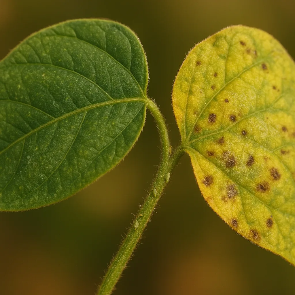
- Rusts (Puccinia, Uromyces spp.). Bright orange, yellow, brown or black pustules (uredinia/urediniospores) typically on leaf undersides; pustules 0.5–2 mm, often in linear or scattered patterns. Common on cereals, legumes, ornamentals; seasonality varies with host but often spring and fall.
- Anthracnose (Colletotrichum spp.). Irregular sunken necrotic lesions on stems, fruit, or leaves; dark brown to black centers with orange or pink spore masses in humid conditions. Frequently affects trees (e.g., Sycamore, oak) and fruit crops.
- Botrytis blight (gray mold) (Botrytis cinerea). Fuzzy, gray to brown conidial masses on soft tissues—flowers, fruit, succulent stems. Lesions often soft, water-soaked initially, then covered by fuzzy spores. Favored by cool, wet periods (10–20°C).
Oomycete diseases (water molds)
- Downy mildew (Peronospora, Plasmopara spp.). Pale, yellow or chlorotic spots on upper leaf surfaces with downy white/gray sporulation beneath. Prefers cool (10–20°C), wet conditions; common on grapevine (Plasmopara viticola), lettuce, brassicas.
- Phytophthora root and crown rot (Phytophthora spp.). Dark brown, water-soaked lesions at crown or roots; blackened, soft roots that easily slough off. Associated with waterlogged soils and heavy clay; widespread in temperate to subtropical regions.
Bacterial diseases
- Bacterial leaf spot (Xanthomonas spp., Pseudomonas syringae). Angular lesions limited by leaf veins, 2–10 mm expanding to larger necrotic patches; often water-soaked margins and translucent halos. Bacteria may ooze a sticky exudate in warm conditions.
- Bacterial canker (Pseudomonas syringae pv. syringae, Clavibacter spp.). Sunken lesions, dieback on stems/branches; gummy sap on pruning cuts. Important in fruit trees and ornamentals.
Viral diseases
- Mosaic viruses (Tobamovirus, Potyvirus). Mottled light/dark green or yellow patches on leaves, distorted growth, stunting. Often transmitted by aphids or mechanical contact; symptoms can be systemic and persistent.
- Chlorotic ringspots. Circular or ring-shaped yellow patterns; size can range from a few millimeters to several centimeters depending on host and virus.
Abiotic disorders (non-infectious)
- Nutrient deficiencies: Nitrogen deficiency—uniform pale green, older leaves first; potassium deficiency—marginal scorch and chlorosis; magnesium deficiency—interveinal chlorosis typically on older leaves.
- Physiological burns and salt damage: Scorched leaf margins, tan or brown tissue, often with crisp texture; localized to areas with salt exposure or fertilizer overdose.
- Environmental stress: Frost damage—water-soaked cells turning black brown; sunscald—white or bleached patches on south-facing trunks or leaves.
Diagnostic workflow: step-by-step plant problem identification (and how Orvik helps)
Follow a repeatable workflow rather than guessing. Use your phone to document symptoms, and consider Orvik’s AI visual tools to speed up initial classification before deeper testing.
For more on this topic, see our guide on Diagnose and Fix Plant Problems Fast.
- Document the problem. Take clear photos of: the entire plant, affected leaves/stems/roots, underside of leaves, and context (soil, nearby plants). Include a ruler or coin for scale; lesion size is diagnostic (e.g., rust pustules 0.5–2 mm).
- Record environment. Note recent weather (days of rain, humidity), irrigation frequency (e.g., daily overhead vs. weekly drip), soil type (sandy, loam, clay), and fertilizers used (N-P-K ratios).
- Observe symptom progression. Are symptoms spreading outward from watering points? Is only new growth affected? Rapid systemic wilting suggests vascular pathogens (Verticillium, Fusarium).
- Use digital tools. Upload high-quality images to Orvik or a lab service. Orvik’s visual AI can triage likely categories (fungal, bacterial, viral, abiotic) and suggest close matches based on image libraries and metadata.
- Simple at-home tests. Smell for musty or sour odors (root rot/Phytophthora); check for slimy or sticky ooze (bacterial). Carefully wash a small root sample to inspect fine root loss or discoloration.
- When to send samples to a lab. If the problem is widespread, affects valuable plants (fruit trees, high-value ornamentals), or appears to be regulated (e.g., suspected Xylella fastidiosa), send samples to a plant diagnostic clinic for culture, PCR, or microscopy.
- Keep records: date-stamped photos and notes help detect patterns and seasonality.
- When using Orvik, provide context: host species, location, recent treatments—AI accuracy improves with metadata.
Houseplant vs. garden disease identification
Houseplants and garden plants share many pathogens, but differences in environment and management change the likely causes and diagnosis approach.
Houseplants: common issues and visual cues
- Root rot (Phytophthora, Pythium): Brown, mushy roots that detach easily; yellowing of lower leaves and collapse of new shoots. Common in poorly draining pots and overwatering.
- Powdery mildew: White dusty coating on philodendron or begonias in dry, crowded indoor conditions—distinct from dust because powder wipes off revealing healthy tissue beneath.
- Spider mite injury (not a disease but often confused): fine stippling, webbing, tiny specks visible with 10x lens; often in dry indoor air.
Garden (outdoor) plants: common issues and cues
- Downy mildew on lettuce or brassicas: yellow angular spots with white/gray downy growth beneath—favored by cool, damp conditions in spring/fall.
- Rust on beans and roses: distinct orange/brown pustules on undersides—more common outdoors where wind spreads spores.
- Soilborne pathogens: Fusarium/Verticillium wilts causing unilateral leaf yellowing, stunting, and vascular discoloration (brown streaks inside stems).
Key differences:
You may also find our article on Identify Plants and Diagnose Problems with AI helpful.
- Indoor plants: suspect cultural/abiotic problems and root pathogens from overwatering; pests like mealybugs and spider mites are frequent.
- Outdoor plants: airborne fungal pathogens (rusts, mildews), soilborne oomycetes, and bacterial infections spread by rain and splashing are more common.
Comparisons: How to tell lookalikes apart
Misidentifying lookalike symptoms wastes time. Below are side-by-side comparisons with visual and contextual tests to tell them apart.
Powdery mildew vs. Downy mildew
- Powdery mildew: white, talc-like coating on upper leaf surfaces; spores form in dry, warm conditions. Wiping the surface often removes powder.
- Downy mildew: pale or yellow angular lesions on upper surface with fuzzy, downy sporulation on the underside; thrives in cool, wet conditions and cannot be wiped off easily.
- Diagnostic test: check leaf underside for sporulation and note weather history (wet/cool = downy; dry/warm = powdery).
Fungal leaf spot vs. Bacterial leaf spot
- Fungal spots (Colletotrichum, Alternaria): often round to irregular with concentric rings or target-like patterns; spores may be visible as powdery masses under magnification.
- Bacterial spots (Xanthomonas, Pseudomonas): angular lesions often bounded by veins, water-soaked margins, and sometimes a yellow halo. Bacterial ooze may appear on warm, wet days.
- Diagnostic test: place a small infected leaf in a sealed plastic bag for 48 hours—bacterial lesions may produce translucent exudate; culture or lab tests confirm bacteria.
Nutrient deficiency vs. disease
- Nutrient deficiency: symptoms follow predictable patterns (e.g., interveinal chlorosis for Mg deficiency), often symmetrical across similar-aged leaves, and not associated with signs like fungal spores or lesions.
- Disease: often asymmetrical, with discrete lesions, necrosis, or sporulation. Rapid spread or presence of pests increases disease likelihood.
- Diagnostic test: check soil pH and perform a simple tissue or soil nutrient test; correctable deficiencies often respond to balanced fertilization in 7–21 days.
Pest damage vs. disease
- Pests (aphids, caterpillars): look for chewing marks, frass, or insects on stems/undersides. Damage is often localized and irregular.
- Disease: lesions, wilting, or vascular discoloration without insect presence. Secondary pests may colonize diseased tissue, so inspect carefully.
Treatment, prevention and safety
Treatment should be targeted to the identified cause. Follow integrated pest management (IPM) principles—cultural controls first, then biologicals, then chemical controls as needed.
You might also be interested in Mastering Visual ID: Your Photo Identifier Guide.
Cultural controls (first line)
- Improve air circulation: space plants to achieve >30 cm between canopy surfaces in densely planted beds.
- Avoid overhead watering: use drip or soaker hoses to reduce leaf wetness period from 12+ hours (which favors many fungal pathogens).
- Sanitation: remove and destroy heavily infected plant parts; do not compost material infected by systemic pathogens (Verticillium, Phytophthora).
- Use well-draining media for containers: mix potting soil with 10–30% coarse sand or perlite for susceptible houseplants to prevent waterlogging.
Biological and chemical options
- Biologicals: Bacillus subtilis–based products (e.g., strain QST 713) provide preventative suppression of foliar fungi; Trichoderma spp. can suppress soilborne pathogens when applied to potting mixes.
- Chemicals: fungicides with active ingredients like azoxystrobin, pyraclostrobin, or chlorothalonil for foliar control; use copper or mancozeb for some bacterial/fungal complexes. Follow label rates—do not exceed recommended ppm or application frequency.
- Systemic options: for vascular wilts, there are no effective curative sprays—use resistant varieties and crop rotation instead.
Safety and toxicity warnings
- Always read and follow label directions. Many fungicides and bactericides are toxic to aquatic life—avoid application near waterways.
- Wear PPE (gloves, eye protection) when handling concentrated chemicals. Store chemicals away from children and pets.
- Disposal: double-bag and remove infected plant material from site if suspecting regulated pathogens (consult local extension services). Do not burn diseased plant material; some spores spread via smoke.
- Food safety: for edible crops, observe pre-harvest intervals (PHI) listed on product labels.
Prevention and long-term control
Prevention reduces repeated outbreaks and long-term soil inoculum. Adopt practices that lower inoculum levels and reduce plant stress.
Related reading: Which plant is the true money plant?.
- Choose resistant cultivars: e.g., tomato varieties with Fusarium (F) and Verticillium (V) resistance carry genes like Ve and I.
- Crop rotation: rotate non-host crops for 2–4 years for soilborne pathogens when practical.
- Soil amendments: solarization (6–8 weeks of 45–60°C soil temperatures under clear plastic) can reduce some soilborne pathogens in warm months.
- Regular monitoring: inspect plants weekly during high-risk seasons (spring and fall for many foliar pathogens).
- Maintain plant vigor: balanced fertilization (follow soil test results), correct pH (most vegetables prefer pH 6.0–7.0), and good watering practices lower susceptibility.
Conclusion
Plant disease identification combines careful observation, knowledge of pathogen biology, and a practical workflow. Start with clear photographs and environmental notes, triage with tools like Orvik to narrow likely causes, and use cultural controls and targeted treatments. Accurate ID saves time and resources—preventive practices and careful sanitation are your best long-term defenses. When in doubt, collect samples and consult a diagnostic lab or extension service for confirmation.
Orvik’s visual identification can speed the triage process by suggesting probable pathogen categories based on images and metadata; use it as a complement to hands-on inspection and lab tests for the best outcomes.
Frequently Asked Questions
- What is the first thing I should do when I notice symptoms?
- Document the problem with clear photos (whole plant, affected parts, undersides of leaves) and note recent weather, watering, and fertilization. This information guides diagnosis and improves AI or lab assessments.
- Can an app like Orvik reliably identify plant diseases?
- Apps like Orvik provide fast, data-driven triage to narrow likely causes (fungal, bacterial, viral, abiotic). They speed up diagnosis but are best used with environmental context and, when necessary, lab confirmation.
- How can I tell if a spot is fungal or bacterial?
- Fungal spots often have concentric rings, powdery spores, or fuzzy growth; bacterial spots are frequently angular (bounded by veins), water-soaked, and may exude sticky ooze. Lab culture/PCR gives definitive results.
- When should I send a sample to a lab?
- Send samples when the plant is valuable, the disease spreads rapidly, symptoms suggest a regulated pathogen, or when treatments have failed. Labs will perform cultures, microscopy, or PCR for precise ID.
- Are there safe home treatments for common fungal diseases?
- Cultural controls—improved air circulation, reduced leaf wetness, and sanitation—are the safest first steps. Biological products (Bacillus, Trichoderma) are low-risk. Apply chemical fungicides only per label instructions if needed.
- How do I prevent root rot in houseplants?
- Use well-draining potting mixes, containers with drainage holes, water only when top 2–3 cm of soil are dry, and avoid waterlogged saucers. Repot every 1–2 years and inspect roots for firmness and color.
- Can nutrient deficiencies mimic diseases?
- Yes. Deficiencies often produce symmetrical, predictable patterns (e.g., interveinal chlorosis for Mg deficiency) while diseases cause discrete lesions, necrosis, or sporulation. Soil and tissue tests help distinguish them.